Apicoectomy
Microsurgical root-end treatment
When an infection persists in the bone around the root tip even after a standard root canal or retreatment, a microsurgical apicoectomy may be required. Also known as root-end resection, this procedure accesses the root tip surgically through a small gum incision, removes the infected tip along with surrounding infected tissue, and places a small filling to seal the root end permanently.
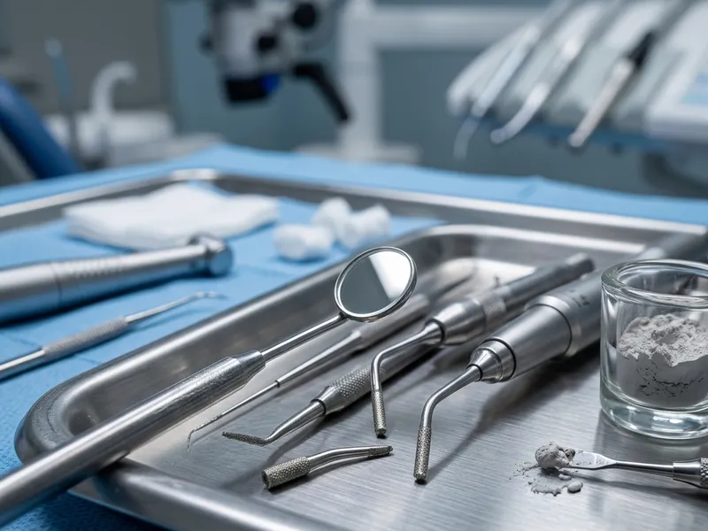
Precision surgical endodontics
An apicoectomy is a delicate surgical procedure. At Shwetha Dentistry, our specialists perform it under high magnification using microsurgical ultrasonic tips and biocompatible retro-filling materials such as MTA or bioceramics.
Gum-level access
Direct access to the root tip, preserving your existing dental crown.
Root-end resection
Removing the infected root tip (apical 3 mm) where bacteria hide.
Retro-filling seal
Sealing the root end with biocompatible materials to prevent leakage.
Who is it for?
- Patients with persistent bone infections after root canal retreatment
- Teeth with calcified canals that cannot be accessed through the crown
- Failed root canals with complex root anatomy or blockages at the tip
- Cases with cysts or infected tissue around the root apex
What to expect
This is a minor surgical procedure performed in our dental chair. The area is completely numbed using local anaesthesia, so you feel no pain. Our team works gently and uses fine sutures to close the gum tissue afterwards. Post-operative swelling and mild soreness are normal for 2–3 days.
Performed at Shwetha Dentistry by our specialist endodontic team.
The Procedure, Step by Step
Exactly what happens during your apicoectomy — no surprises.
Gum Incision & Exposure
Gum Incision & Exposure
Making a small incision in the gum to expose the bone window.
Local anaesthesia is administered to ensure complete comfort. The surgeon makes a small, precise incision in the gum tissue near the tooth to expose the underlying bone and locate the root end.
Gum-level access preserves the existing crown on the tooth.
Bone Window Cavity
Bone Window Cavity
Creating a tiny window in the bone to reach the root tip.
Using specialised microsurgical instruments, the surgeon creates a very small window in the bone to access the infected area and root tip directly. Infected granulation tissue is removed.
The window is kept as small as possible to promote rapid bone healing.
Root-End Resection
Root-End Resection
Removing the infected root tip (apical 3 mm).
Under the microscope, the surgeon resects the apical 3 mm of the root tip — the area containing complex micro-anatomy and delta-like branches where bacteria hide.
Removing the tip eliminates the primary source of the persistent infection.
Ultrasonic Retro-Preparation
Ultrasonic Retro-Preparation
Preparing a small cavity at the resected root end.
Using specialised micro-ultrasonic tips, the surgeon prepares a small, 3 mm deep cavity at the resected end of the root, cleaning the canal opening from the bottom up.
Ultrasonic preparation ensures clean, flat walls for sealing.
Retro-Filling & Suturing
Retro-Filling & Suturing
Sealing the root end and suturing the gum tissue.
The micro-cavity is filled with a biocompatible retro-filling material (such as MTA or bioceramics) to seal the canal permanently. The gum flap is repositioned and closed with fine sutures.
The sutures hold the gum in place for rapid healing and are removed in 5–7 days.
Treatment Cost
₹8,000 – ₹12,000
1 visit (60–90 minutes)
Indicative range — please consult the dentist; the exact price varies from case to case.
Aftercare
- Apply an ice pack to the cheek for 10–15 minutes at a time during the first 24 hours to reduce swelling.
- Eat soft, cool foods and avoid hot, spicy or crunchy foods for a few days.
- Brush your other teeth normally, but avoid brushing directly over the surgical area for the first week.
- Rinse gently with warm salt water after 24 hours. Do not spit forcefully or use straws.
Frequently Asked Questions
Bacteria sometimes hide in the tiny, complex branches at the very tip of the root. If standard treatment cannot reach or sterilise these branches, surgery is needed to remove the tip directly.
The procedure is performed under local anaesthesia, so you feel no pain during surgery. Afterwards, mild soreness and swelling are easily managed.
The gum heals quickly, with sutures removed in 5 to 7 days. Most patients return to normal activities within 24 to 48 hours, though complete bone healing takes a few months.
When performed by a specialist using a surgical microscope and bioceramic materials, the success rate is over 85% to 90%, successfully saving the tooth.
It ranges from ₹8,000 to ₹12,000 — a specialised micro-surgical procedure requiring surgical equipment, suturing materials and biocompatible cements. Please consult the dentist for an exact quote.
No. Because the root is accessed through the gum and bone, the existing crown is left completely untouched and does not need replacing.
Retreatment accesses the canal through the crown of the tooth to replace old fillings. An apicoectomy accesses the root tip surgically through the gums to remove the infected tip.
If surgery fails to resolve the bone infection, the tooth will likely need to be extracted and replaced with a dental implant or bridge.
No. The incision is made carefully, and once healed, the gum tissue returns to its normal appearance with no visible scarring.
Yes, but it is most commonly performed on front teeth, premolars and the front roots of molars due to surgical accessibility.